Fighting Cancer with 3D Printing


Latest News
January 21, 2015
We’ve written in the past about 3D printing solutions that help surgeons better plan complex procedures. According to a Reuters report, British researchers have now created replica models of cancerous tumors and organs that can help doctors target their treatments more precisely.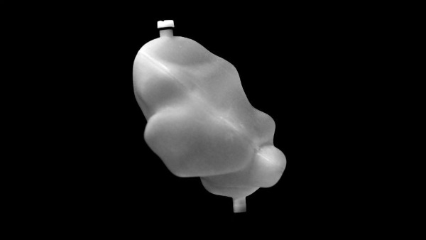
Originally, researchers at the Institute of Cancer Research in London built hand-made models of organs to model radiotherapy treatments. 3D printing has made those tests much more reliable and precise.
The printed tumors and organs, generated with CT scans, can be filled with liquid that allows physicians to see the flow of radiopharmaceuticals, the radioactive drugs that are used to kill cancer cells. Using the models, doctors can more accurately determine the right dosage to use to kill the cancer cells without damaging the surrounding tissues.
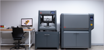
According to the researchers, these tumor “phantoms,” as they are referred to, were made from liver, spleen and kidney scans using a Stratasys printer.
“Each organ was converted to a shell and filling holes and leg supports were added using computer aided design software and prepared for printing,” the researchers wrote. “Additional fixtures were added to the liver to allow lesion inserts to be fixed within the structure. Phantoms were printed from an ultraviolet curable photopolymer using polyjet technology on an Objet EDEN 500V 3D printer. Results: The final print material is a clear solid acrylic plastic which is watertight, rigid, and sufficiently durable to withstand multiple assembly and scanning protocols.”
“The big challenge we faced was to produce a model that was both anatomically accurate and allowed us to monitor the dose of radiation it received,” said Dr. Jonathan Gear, Clinical Scientist at The Institute of Cancer Research. “We found that the printed replicas could give us information we couldn’t get from 2D scans, you will always get more information from a 3D model than a flat image.”
Tests at the Institute and at the Royal Marsden NHS Foundation Trust found that the models could help accurately calculate the required dosage of the radiopharmaceuticals.
Glenn Flux, head of radioisotope physics at the facility, and his colleagues published a paper on the process in Medical Physics over the summer. The organ geometry of the models showed close correspondence to anatomical references. You can read the abstract here.
Source: Reuters
Subscribe to our FREE magazine, FREE email newsletters or both!
Latest News
About the Author
Brian Albright is the editorial director of Digital Engineering. Contact him at [email protected].
Follow DE