Successful 3D Cartilage Printing
Formation of blood vessels


June 19, 2017
Researchers at Belgium’s Sahlgrenska Academy and Chalmers University of Technology have 3D bioprinted new cartilage for transplant procedures.
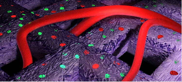
 A model of the formation of blood vessels in the printed cartilage. Image: Chalmers
Scientists have already bioprinted putative cartilage grafts (called autologous chondrocyte implantation) in the past, but existing methods require two surgical procedures and is dependent on the quality and quantity of the patients’ autologous cells, according to the new research.
The research team investigated whether they could use an “established and defined human-derived induced pluripotent stem cell (iPSC) line for bioprinting. This would reduce the need for additional surgeries and provide a larger supply of more controllable cellular material.
The team used iPSCs that originated from chondrocytes and an iPS generation process to rejuvenate cells into the blastula stage. Those cells were then directed to develop into chondrocytes. Essentially, they removed cartilage cells from patients already undergoing knee surgeries, then reverted them back to stem cells.
That material was mixed with nanocellulose hydrogel and bioprinted using hardware from Cellink, then treated with growth stimulants to grow back into cartilage. The new construct was implanted into mice.
The cartilage tissue was able to grow in the animal model, and the researchers noted the formation of blood vessels between the materials.
“What we see after 60 days is something that begins to resemble cartilage. It is white and the human cartilage cells are alive and producing what they are supposed to. We have also been able to stimulate the cartilage cells by adding stem cells, which clearly promoted further cell division,” said Lars Kölby, senior lecturer at Sahlgrenska.
“In nature, the differentiation of stem cells into cartilage is a simple process, but it’s much more complicated to accomplish in a test tube. We’re the first to succeed with it, and we did so without any animal testing whatsoever,” said lead researcher Stina Simonsson.
The research was printed in Scientific Reports.
However, the cellulose used in the current procedure may not be suited for use in humans. The researchers are now looking for another material that could be broken down after implantation and absorbed.
Source: CBS News
Subscribe to our FREE magazine, FREE email newsletters or both!
About the Author
Brian Albright is the editorial director of Digital Engineering. Contact him at [email protected].
Follow DE