Helping design and engineering professionals discover, evaluate and specify technologies and processes that shorten the design cycle and enable success.
Alert!
Digital Engineering ceased publication on July 1, 2026. This website remains available as an archive of engineering content.
For inquiries or information, please email [email protected].

Digital Engineering April 2026
In the latest issue of Digital Engineering, we take a look at the latest innovations in design for additive manufacturing, including the use of natural language inputs, social media cosplayers, and AI integration. The issue also includes a feature…

January Special Focus Issue: Design for Additive
In this Special Focus Issue of Digital Engineering, learn about the latest advancements in design for additive manufacturing, including new software tools, additive in automotive, custom medical devices, and more.
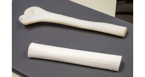
Researchers at University of Texas Southwestern Medical Center have developed a 3D printing technique for generating realistic models of the human femur that could simplifiy the ability to conduct biomechanical research.
The study, published in the Journal of Orthopaedic Research in collaboration with researchers at The University of Texas at Dallas, is validating the use in biomechanical studies of femurs produced using cost-efficient 3D printers and materials, according to researchers. Though the study focused on replicating the femur (thigh bone) and its mechanical properties, the process could be used ito build models of any human bone for research.
“The femur is a focal point of biomechanical research because of its critical role in weight-bearing and mobility,” says lead author Robert Weinschenk, MD, assistant professor of Orthopaedic Surgery and Biomedical Engineering at UT Southwestern. “Traditionally, biomechanical researchers have used cadaver or synthetic bones for studies, but those can be expensive, difficult to obtain, and have inherent limitations. The use of 3D printing to generate humanlike bones can be a significant boost to researchers studying new surgical techniques and conditions such as osteoporosis, traumatic fractures, deformities, and benign or malignant bone lesions.”
Collaborating with mechanical engineers from UT Dallas, Dr. Weinschenk and the team used polylactic acid—a biodegradable polyester material commonly used in 3D printing—to construct a range of femur models with different physical attributes such as wall thickness and infill density. Those models were then tested for flexural strength using three-point bending, and results were compared to the biomechanical response of human femurs, enabling the team to identify the methodology that produced the most accurate replica.
Kishore Mysore Nagaraja, a Ph.D. candidate at UT Dallas, developed numerous samples of the printed femurs and tested them to ensure they were mechanically equivalent to actual femur bones.
Four generations of synthetic femur models have been developed for biomechanical testing and sold commercially since 1987, according to Dr. Weinschenk. However, they have had limitations, including cost and delivery time. He said the 3D printing technique he and his colleagues created solves those problems.
“We think this is novel and can gain wide use and acceptance because anyone with a cheap 3D printer can download the file, print the specimen, and do their own studies in an inexpensive way without delay,” Dr. Weinschenk says.
Dr. Weinschenk and his UT Southwestern and UT Dallas colleagues made models of the middle portion of the femur, just under 8 inches in size and almost an inch in diameter. The specimens are produced at an estimated cost of $7.
Researchers at UT Dallas focused on the mechanical evaluation and characterization of the 3D-printed femur.
“With 3D printing, we’re able to print out the femur bone with the same geometry of the femur inside the body,” says Dr. Wei Li, Ph.D., Assistant Professor of Mechanical Engineering at UT Dallas and the study’s senior author. “In our biomechanical tests, the femur performed as well as a human femur.”
Sources: Press materials received from the company and additional information gleaned from the company’s website.

DE's editors contribute news and new product announcements to Digital Engineering. Press releases may be sent to them via [email protected].
Follow DE


About Us · Contact Us · Editorial Team · Advertising · Privacy Policy · Subscriber Services · © 2026 Digital Engineering 24/7 · Peerless Media